
Multispectral Optoacoustic Tomography
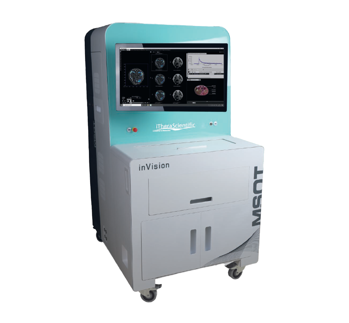
Multispectral Optoacoustic Tomography
Multispectral Optoacoustic Tomography (MSOT) or Photoacoustics utilizes pulsed light at multiple wavelengths and detection of acoustic responses. Images are generated and spectrally unmixed to yield the biodistribution of reporter molecules and tissue biomarkers.
MSOT offers the unique ability to accurately visualize, track and quantify endogenous chromophores, nanoparticles, biomarkers and optical agents, in vivo and in real time. Excellent intrinsic tissue contrast by combining the molecular specificity of optical imaging with the penetration depth and 3D resolution of ultrasound. Offering the best of the Optical and Ultrasound Imaging.
3 Imaging Modes in 1 System
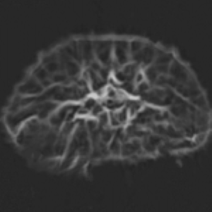
Optoacoustic
• Represents absorbance
• Blood as main component
• Versatile contrast agents
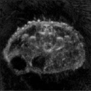
Reflection UCT
• “Classic” ultrasound
• Whole-body view
• Tomographic quality
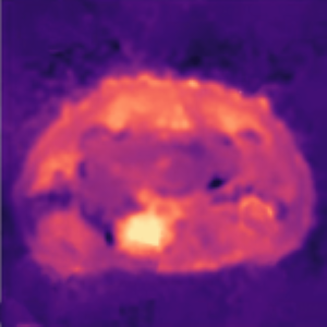
Transmission UCT
• Measures speed of sound
• Indicates tissue stiffness
• Unique new information
Features
- High sensitivity (nmol)
- Real-time image visualisation at video rate
- Volumetric deep-tissue imaging (whole-body penetration in small animal imaging applications)
- Single and multi-wavelength imaging at video rate
- Data post-processing suite for spectral and temporal analysis
- Ultrasound detector array with 64–512 channels
- Tomographic ultrasound
Applications
- Cancer - Analyse parameters of disease progression across a wide range of biological mechanisms
- Cardiovascular - Monitoring blood flow and the beating heart in real time
- Brain - High-resolution imaging of deep brain structures, through intact skin and skull
- Kinetics and Biodistribution - Quantify kinetic processes in real time
Automated and Intuitive Workflow
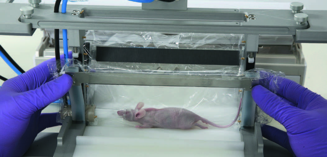
Animal Preparation
• Safe animal handling
• Repeatable fixed position
(key for longitudinal studies)
• Easy mounting of animal in holder
• Integrated anesthesia supply
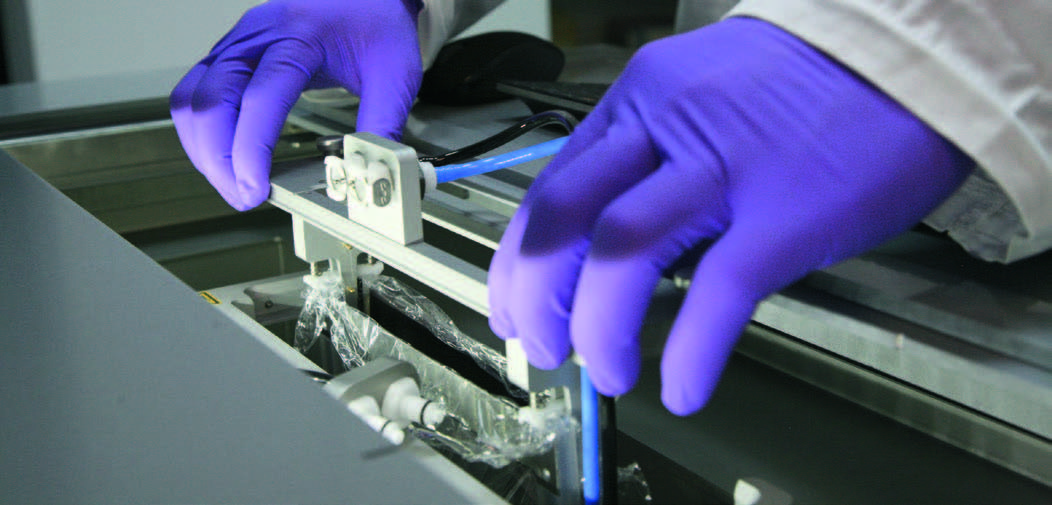
Image Acquisition
• X-sectional scan along the region of interest
• System calibration before scan
• Fully automated image acquisition (OA and US)
• Access for catheter or thermometer
• Maintains animal body temperature
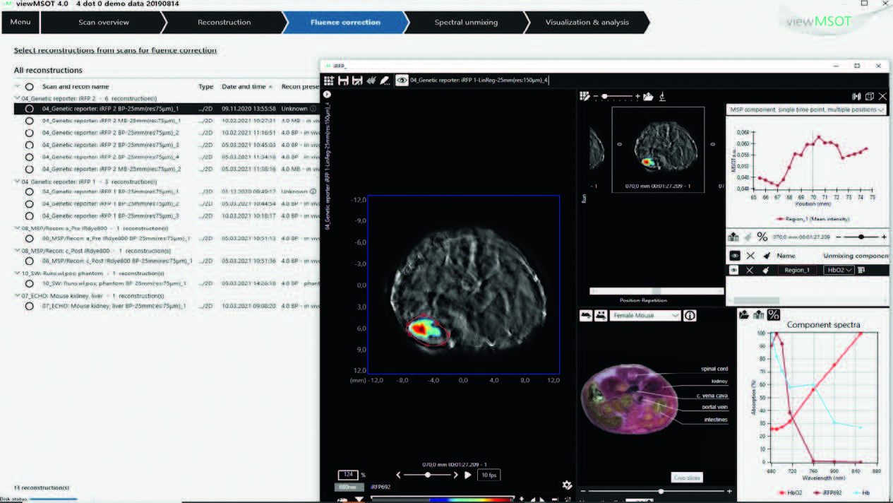
Data Post-Processing
• Quantitative image reconstruction
• Spectral analysis of chromophores
• Visualization and super-positioning
of OA and US images
• Data and image export


