Prospect T1
Small Animal Ultrasound
Ultrasound and Photoacoustic Imaging
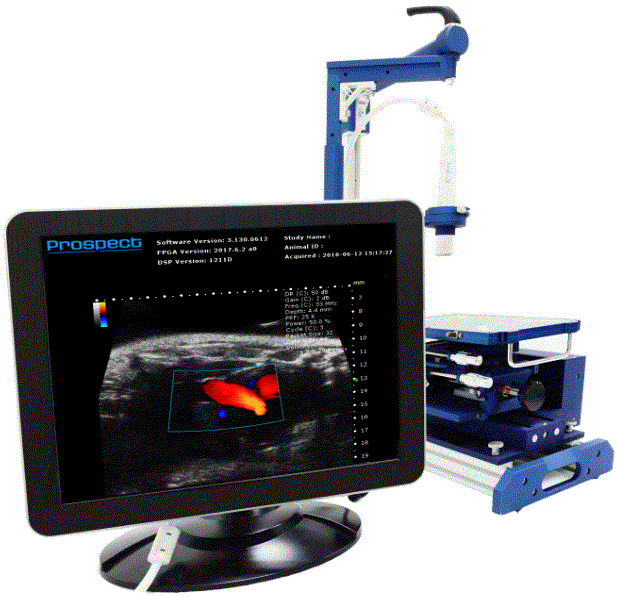
Prospect T1 is a small, economical animal ultrasound imaging system with a spatial resolution down to 30μm. It provides researchers with real-time and non-invasive image acquisition capabilities for in-vivo and longitudinal studies on animal models. With a high frame rate and impressive sensitivity, it is capable of visualising fast moving events such as blood flows and heart motions, as well as micro-vasculatures on mouse models.
Features
-
Real-time color Doppler imaging and triplex imaging in update mode (B/PW/Color).
-
Molecular imaging for perfusion and targeting information.
-
Comprehensive measurement tools for image analysis with 2-D and 3-D image data.
-
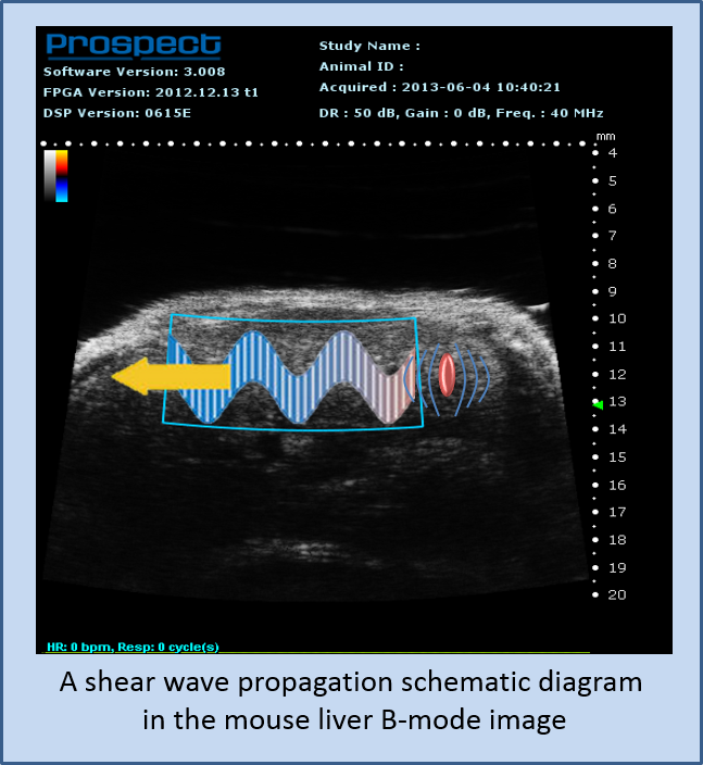
Connectivity to external devices, such as a laser, a HIFU transducer, or a source for generating radiation force, allows extened capabilities including sonoporation and elasticity imaging.
-
Adjustable imaging parameters (center frequency, cycle, PRF, TGC, …etc) for each probe to optimize image quality in different applications.
-
Full line of image adjustments for live viewing and retrospective review.
-
Virtual Array (Synthetic Aperture Focusing Technique) for improved image resolution and depth of field.
-
Save static or dynamic data in multiple data formats including raw, jpg, tif, bmp, DICOM, cineloop (up to 1000 frames) and avi.
Imaging Modes
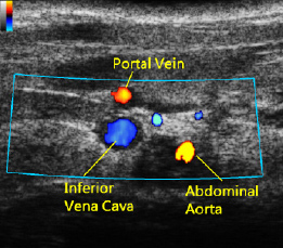
Doppler Mode
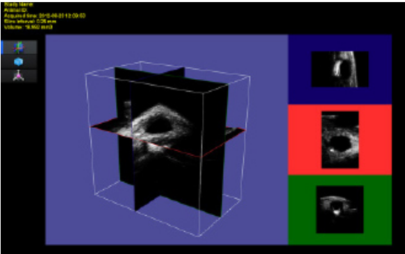
3D Imaging Mode
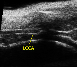
B Mode
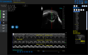
M Mode
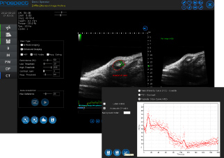
Constrast Mode
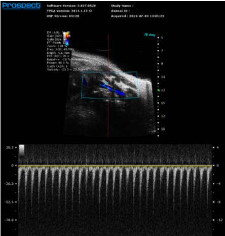
Analog Doppler Mode
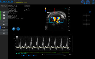
Pulsed-wave (PW) Doppler Mode
